A practical, patient-friendly visual guide

Understand the spots and changes on your skin

Clear, reliable information to help you recognise common skin lesions, understand how doctors assess them, and know when to seek advice.

Clinical + dermoscopic views
Images teach patterns—not self-diagnosis.
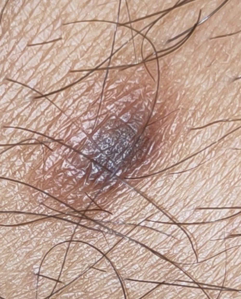
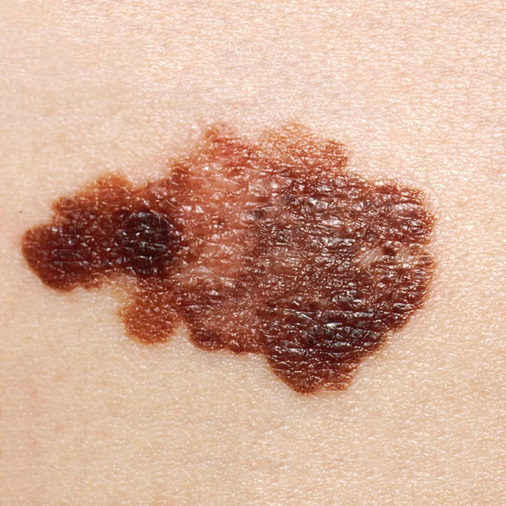
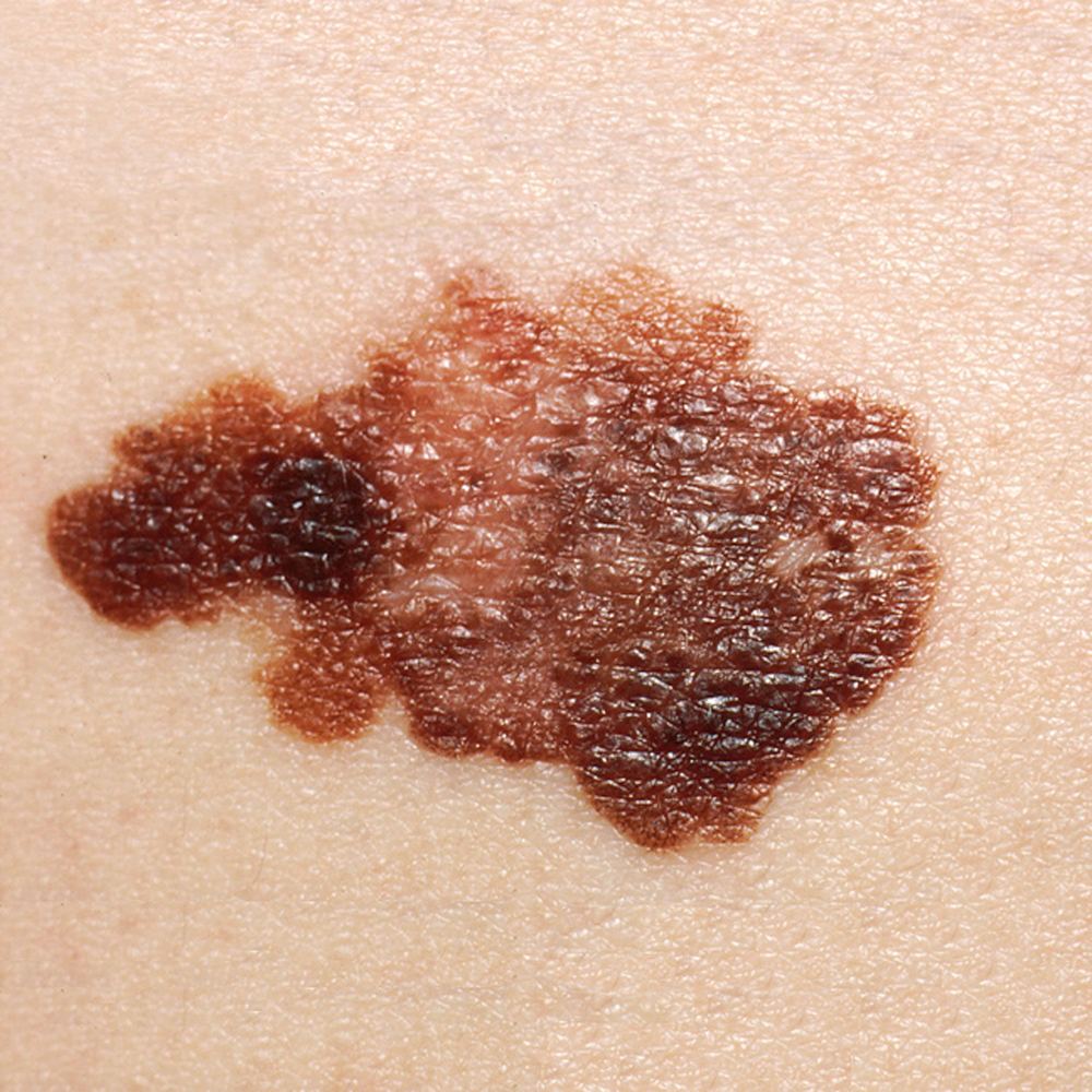
Clinical photograph of a melanoma showing asymmetry and variation in colour
Clinical view
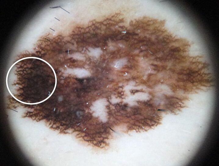
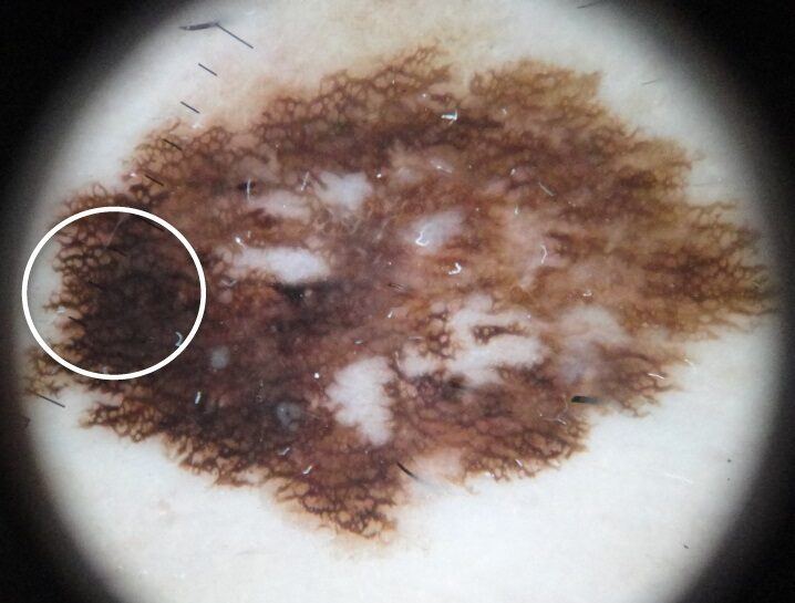
Dermoscopic view of a superficial spreading melanoma in situ
Dermoscopy
Clinical photograph of a basal cell carcinoma
Skin cancer can look different from person to person

Do not wait for every sign

When should I arrange a prompt review?

A new or changing lesion, a sore that will not heal, repeated bleeding or crusting, pain or tenderness, or a spot growing over weeks should be assessed by a qualified clinician.

What to do next

Start with the important three

Common skin cancers

Basal cell carcinoma (BCC), squamous cell carcinoma (SCC) and melanoma are the main skin cancers discussed here. They may resemble harmless spots, and photographs cannot confirm a diagnosis.

Dermoscopy is a magnified examination used by trained clinicians. A biopsy is often needed for certainty.

Explore common skin cancers

Clinical photograph of melanoma with asymmetry, an irregular border and varied pigmentation
Macro clinical photographIn this example: asymmetry, an irregular outline and several shades of brown and black.Example only. Image: National Cancer Institute, public domain.
Dermoscopic photograph of superficial spreading melanoma in situ showing an atypical broadened pigment network
Dermoscopic photographIn this example: an asymmetrical pattern with an atypical, broadened pigment network.Example only. Image: Braga et al., PLOS ONE, CC BY.

What a patient may notice

  • A new spot or an existing mole that changes
  • Asymmetry, irregular border or several colours
  • A firm, raised lesion that is growing
  • Bleeding, itching or ulceration without another explanation

Potentially serious · early review matters

Melanoma

01

A cancer of pigment-producing melanocytes. It is less common than BCC or SCC but is more likely to spread if not found early.

How common?

Melanoma is one of Australia’s most commonly diagnosed cancers. Risk rises with age, but it can occur in younger adults.

Main causes and risks

UV exposure—especially episodes of sunburn—is the major preventable risk. Fair skin, many or atypical moles, family or personal history, and immune suppression also increase risk. Melanoma can occur on skin with little sun exposure.

Main clinical types

Superficial spreading melanoma commonly enlarges across the skin before invading more deeply. Nodular melanoma often presents as a firm, rapidly enlarging lump and may not satisfy the ABCDE rule. Lentigo maligna melanoma usually develops on chronically sun-damaged facial skin in older adults. Acral lentiginous melanoma occurs on palms, soles or beneath nails and is relatively more important in darker skin types. Amelanotic melanoma may be pink, red or skin-coloured.

What is melanoma in situ?

Melanoma in situ (Stage 0 melanoma) means abnormal melanocytes are confined to the epidermis—the outer layer of skin—and have not invaded the dermis. It often appears as a slowly enlarging, irregular flat patch with uneven colour; lentigo maligna is an in-situ subtype usually found on chronically sun-damaged facial skin. Pathology confirms the diagnosis. Because there is no dermal invasion, Breslow thickness is not assigned and the lesion itself cannot spread to lymph nodes or distant organs. Complete surgical removal is usually curative, commonly with a further margin after the diagnostic biopsy. Large or poorly defined facial lesions may need staged margin-controlled surgery. Untreated lesions can enlarge and some eventually develop an invasive focus, so treatment and ongoing skin surveillance remain important.

What is desmoplastic melanoma?

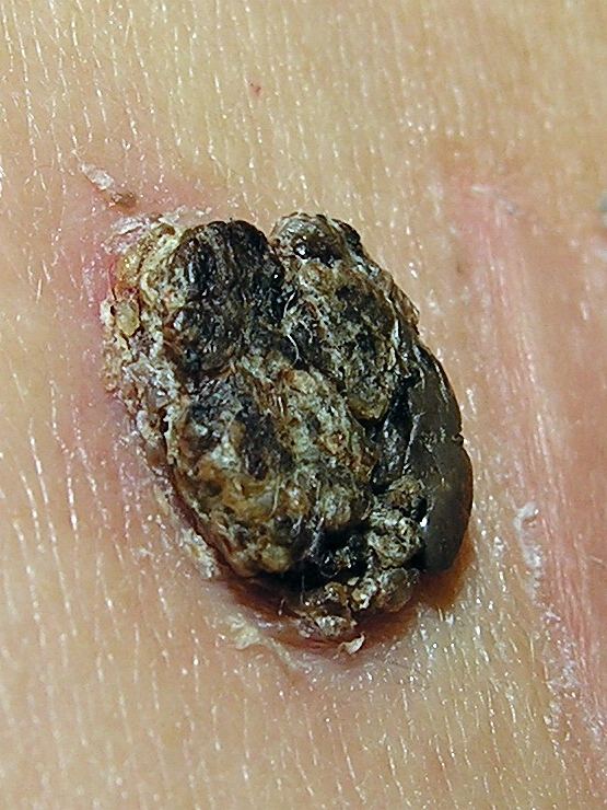
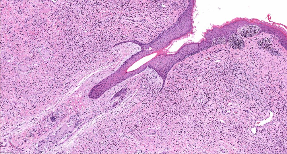
Desmoplastic melanoma is a rare invasive melanoma in which malignant spindle-shaped melanocytes lie within dense fibrous tissue. It usually affects older, fair-skinned people on chronically sun-damaged head or neck skin and may arise beneath lentigo maligna. It may look like a firm, pale, pink or scar-like thickening rather than a dark mole, making recognition and partial-biopsy diagnosis difficult. Involvement of small nerves (neurotropism) can cause pain, tingling or numbness and increases concern for local recurrence. Diagnosis requires an adequately deep biopsy and specialist dermatopathology. Wide surgical excision is the main treatment; margin-controlled surgery, radiotherapy and staging investigations are considered according to site, thickness, margins, neurotropism and whether pathology describes a pure or mixed subtype. Management should be planned by an experienced melanoma team.

Appearance and usual sites

Melanoma may be a new lesion or a changing existing mole. Warning patterns include asymmetry, an irregular edge, uneven colour, evolution, or a spot unlike the person’s other lesions. Men more often develop melanoma on the trunk and women on the legs, but it can arise anywhere—including scalp, nails, soles, genital skin and rarely the eye or mucosa.

What may be seen with dermoscopy?

A trained clinician may see an atypical or broadened pigment network, irregular dots or globules, streaks, blue-white structures, regression, asymmetrical structureless areas or atypical blood vessels. No single feature proves melanoma; the complete pattern and clinical history matter.

What else can resemble it?

Atypical or irritated moles, seborrhoeic keratosis, pigmented BCC, dermatofibroma, a blood blister and benign nail pigmentation may resemble melanoma. Conversely, melanoma can look deceptively harmless, so persistent change should be examined rather than matched to photographs.

How is it diagnosed?

Assessment includes history, examination of the lesion and surrounding skin, comparison with other moles and usually dermoscopy. When feasible, the entire suspicious lesion is removed with a narrow margin for pathology. The report records features such as melanoma thickness and ulceration. A wider excision is then planned; sentinel lymph-node biopsy and scans are considered only for selected stages.

Treatment options

Surgery is the main treatment for localised melanoma: diagnostic excision is followed by a wider excision based on pathology. Selected patients may be offered sentinel-node biopsy. Higher-risk or advanced melanoma may require specialist immunotherapy, targeted therapy when an actionable mutation is present, radiotherapy in selected situations, or a combination planned by a multidisciplinary team.

Overall outlook

Outlook is excellent for many thin, early melanomas. It becomes less favourable once melanoma is thicker or has spread, which is why prompt assessment of change matters.

After treatment

Follow-up is individualised according to stage and personal risk. It commonly includes scar and lymph-node review, full-skin examinations, self-check education and lifelong sun protection. A previous melanoma increases the chance of another melanoma and other skin cancers.
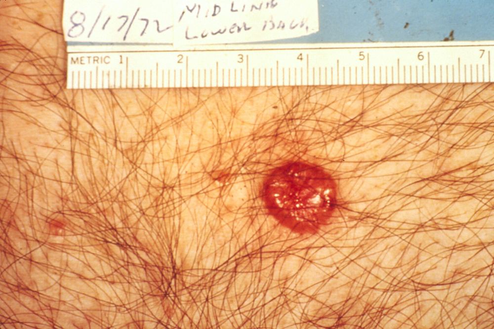
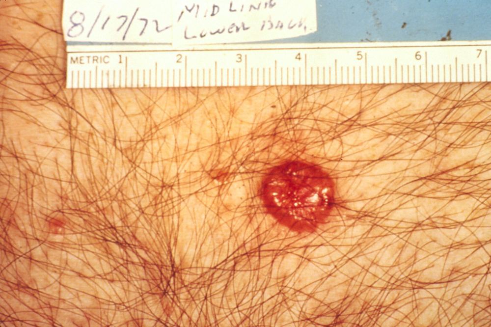
Clinical photograph of a red shiny basal cell carcinoma with an ulcerated centre
Macro clinical photographIn this example: a shiny pink-red lesion with a rolled edge and central ulceration.Example only. Image: National Cancer Institute, public domain.
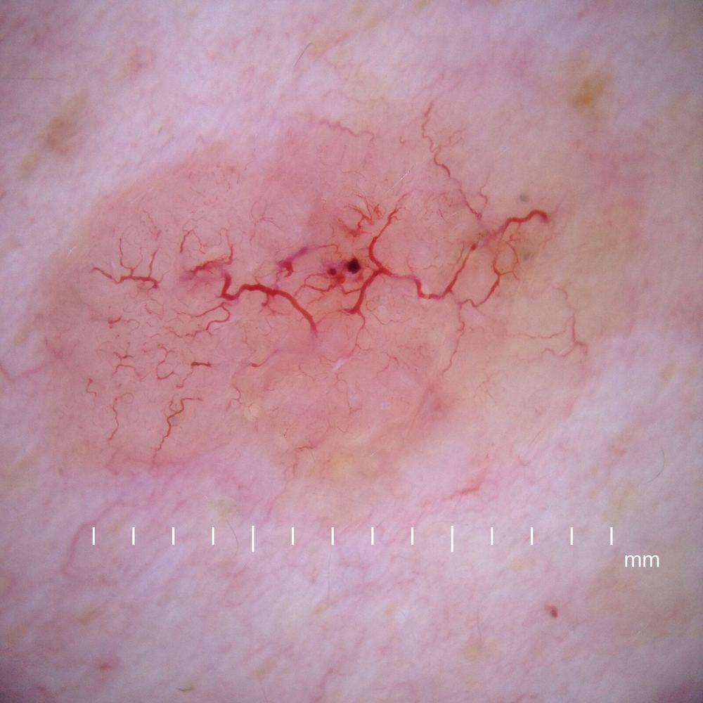
Dermoscopic view of a nodular basal cell carcinoma showing branching blood vessels
Dermoscopic photographIn this example: branching surface vessels and shiny white-red structureless areas.Example only. Image: David Moreno, CC BY-SA.

What a patient may notice

  • A shiny, pearly or translucent bump
  • A sore that heals and returns
  • Repeated bleeding or crusting
  • A red scaly patch or scar-like area that slowly enlarges

Most common · usually slow-growing

Basal cell carcinoma (BCC)

02

A locally invasive cancer arising from basal cells. BCC rarely spreads to distant organs, but untreated lesions can damage nearby skin and deeper structures.

How common?

BCC is the most common skin cancer. It is particularly frequent in Australia and becomes more likely with cumulative UV exposure and age.

Main causes and risks

Long-term and intermittent UV exposure are major contributors. Fair skin, previous BCC, outdoor work, radiation exposure, immune suppression and some inherited syndromes increase risk.

Main clinical types

Nodular BCC is often a pearly or shiny papule with fine surface vessels and may ulcerate. Superficial BCC resembles a slowly enlarging red scaly patch, commonly on the trunk. Infiltrative and morphoeic BCC may be subtle, firm or scar-like with poorly defined extensions. Micronodular and basosquamous patterns can behave more aggressively. Pigmented BCC contains brown, blue or black colour and may mimic melanoma.

Appearance and usual sites

BCC commonly develops on the head, neck and other sun-exposed skin, but superficial BCC often occurs on the trunk. It may bleed after minor contact, crust, appear to heal and then return, or slowly enlarge over months or years. Borders can extend beyond what is visible, particularly with infiltrative subtypes.

What may be seen with dermoscopy?

Typical clues include branching ‘tree-like’ vessels, shiny white-red areas, small erosions or ulceration, blue-grey ovoid nests and leaf-like or spoke-wheel pigmented structures. Superficial lesions may show many fine short vessels. These features guide assessment but pathology confirms the diagnosis when required.

What else can resemble it?

Dermatitis, psoriasis, an irritated seborrhoeic keratosis, scar tissue, a benign mole, sebaceous hyperplasia and melanoma can resemble BCC. A persistent solitary patch or sore deserves assessment even when it causes no symptoms.

How is it diagnosed?

A clinician assesses the border, site, size, recurrence status and dermoscopic pattern. A shave, punch or excision biopsy may be used depending on the lesion and proposed treatment. Pathology identifies the subtype and whether tumour reaches the specimen edges; these details help classify the lesion as lower or higher risk.

How is treatment chosen?

Choice depends on subtype, size, anatomical site, border definition, whether it has recurred, patient health and the desired balance between cure, function and appearance. Facial ‘H-zone’ lesions, aggressive histology, nerve symptoms, recurrence and poorly defined borders generally require more margin-controlled planning.

Treatment options

Standard excision is common. Mohs micrographic surgery offers staged margin examination for selected high-risk, recurrent or tissue-critical lesions. Curettage with cautery may suit selected low-risk lesions. Topical imiquimod or 5-fluorouracil, photodynamic therapy or cryotherapy may be considered for selected superficial disease; radiotherapy can be useful when surgery is unsuitable. Locally advanced or metastatic BCC needs specialist systemic treatment.

Overall outlook

Most BCCs are cured, especially when treated early. Recurrence risk depends on subtype, site, size, borders and previous treatment. People who develop one BCC have a higher chance of developing another.

After treatment

The wound and pathology result should be reviewed, including whether margins are clear. Ongoing skin checks and self-examination are important because both recurrence and new primary skin cancers can occur. A changing scar, new lump, ulcer or persistent numbness near the site should be reported.

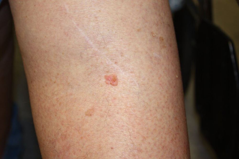
Clinical photograph of a small pink raised squamous cell carcinoma on the leg
Macro clinical photographIn this example: a raised pink lesion with surface scale and keratin.Example only. Image: National Cancer Institute, public domain.
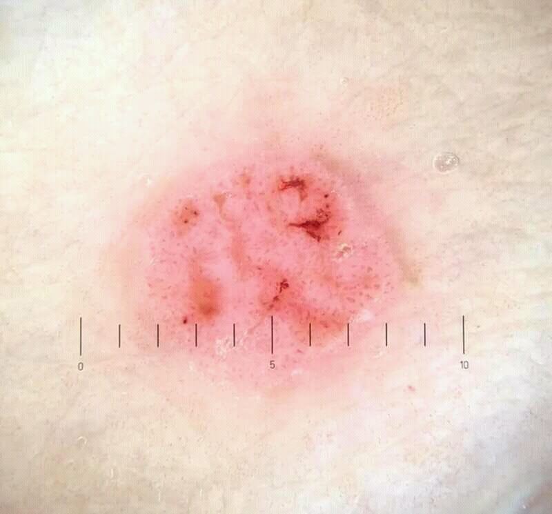
Dermoscopic photograph of squamous cell carcinoma in situ, also called Bowen disease
Dermoscopic photographThis in-situ example shows scale with clustered coiled or glomerular vessels.Example only. Image: Javed M, Wikimedia Commons, CC BY-SA.

What a patient may notice

  • A firm, tender or rapidly growing lump
  • A thick, red, scaly or crusted patch
  • A crater-like growth or persistent ulcer
  • A lesion on the lip or ear, or in a scar, that changes

Common · can sometimes spread

Squamous cell carcinoma (SCC)

03

A cancer of keratin-producing squamous cells. SCC often develops on chronically sun-damaged skin and may grow more quickly than BCC.

How common?

SCC is the second most common skin cancer. It is common on sun-exposed skin in older adults, but risk is strongly influenced by cumulative UV exposure and immune status.

Main causes and risks

Cumulative UV damage is the leading cause. Actinic keratoses, immune suppression, chronic wounds or scars, previous radiation, and some chemical or viral exposures can increase risk.

Main clinical types

SCC in situ (Bowen disease) remains within the epidermis and usually appears as a persistent scaly plaque. Invasive SCC extends into the dermis and may be keratotic, ulcerated or tender. Keratoacanthoma-type SCC can form a rapidly growing crater-like nodule. Less common variants include acantholytic, spindle-cell and desmoplastic SCC, which require pathology assessment.

Appearance and usual sites

SCC usually occurs on chronically sun-exposed areas such as the face, bald scalp, ears, lower lip, forearms and backs of the hands. It may appear as a firm tender lump, thick scaly plaque, cutaneous horn, rapidly growing crater or non-healing ulcer. SCC may also develop in a chronic wound, burn scar, genital skin or an area of previous radiation.

What may be seen with dermoscopy?

Possible clues include surface scale or keratin, white circles, white structureless areas, blood spots, ulceration and irregular coiled, dotted, hairpin or polymorphous vessels. Bowen disease often shows clustered glomerular vessels. Dermoscopy cannot reliably determine invasion depth, so adequate biopsy is important.

What else can resemble it?

Actinic keratosis, inflamed seborrhoeic keratosis, wart, eczema, psoriasis, keratoacanthoma, BCC and amelanotic melanoma can look similar. Pain or tenderness, rapid growth, increasing thickness and persistent ulceration raise concern but are not diagnostic by themselves.

How is it diagnosed?

Assessment includes lesion size and depth, anatomical site, growth rate, nerve symptoms, lymph nodes and immune status. A biopsy must be sufficiently deep to distinguish in-situ from invasive disease and to identify differentiation, thickness, perineural invasion and other high-risk features. Imaging or lymph-node investigation is reserved for selected high-risk disease.

How is treatment chosen?

In-situ and invasive SCC are treated differently. For invasive disease, clinicians consider tumour diameter and thickness, differentiation, site, recurrence, immune suppression, nerve involvement and whether the lesion arose in a scar or chronic wound. High-risk lesions merit specialist or multidisciplinary planning.

Treatment options

Excision is the usual treatment for invasive SCC. Mohs surgery may be appropriate for selected high-risk, recurrent or anatomically critical tumours. Curettage and cautery, cryotherapy, topical treatment or photodynamic therapy may suit selected SCC in situ or carefully chosen low-risk lesions, but are not interchangeable with treatment of higher-risk invasive SCC. Radiotherapy may be primary or additional treatment; nodal or advanced disease may require surgery, radiotherapy and/or immunotherapy.

Overall outlook

Most SCCs are successfully treated. Risk of recurrence or spread is higher for thick, large, rapidly growing, recurrent or poorly differentiated tumours and those on certain sites or in immunosuppressed patients.

After treatment

Follow-up frequency depends on tumour risk and patient factors. Reviews may include the scar, surrounding skin and regional lymph nodes as well as a total-skin examination. New pain, numbness, a lump near the scar or in nearby lymph-node areas, or rapid local change should prompt review.

Sun-damage precursor lesion

Actinic keratosis

Actinic keratosis (AK), also called solar keratosis, is an area of abnormal sun-damaged keratinocytes. It is not yet an invasive skin cancer, but it marks significantly sun-damaged skin and a minority of lesions can progress to squamous cell carcinoma.

A thick, tender, rapidly growing, ulcerated or repeatedly bleeding lesion should be assessed promptly and may require biopsy.

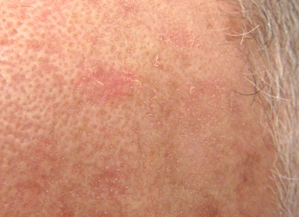
Close clinical photograph of a rough scaly actinic keratosis
Clinical photographA rough, adherent scaly patch on sun-damaged skin. AK may be easier to feel than see.Future FamDoc, Wikimedia Commons
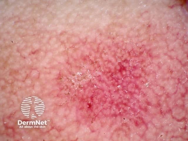
Dermoscopic photograph of facial actinic keratosis showing a strawberry pattern
Dermoscopic photographFacial AK may show a red pseudonetwork around follicles—the “strawberry pattern”—with surface scale.DermNet image licence

How common and why?

AK is very common in Australia, especially in fair-skinned adults with years of cumulative ultraviolet exposure. Risk is higher with outdoor work, bald scalp, advancing age, previous skin cancer and immune suppression. Typical sites are the face, ears, bald scalp, forearms and backs of the hands.

What can it look like?

A small rough or gritty patch that may be skin-coloured, pink, red or brown. It can be flat or thick, scaly, crusted, tender or form a cutaneous horn. Variants include hypertrophic, pigmented, lichenoid and actinic cheilitis affecting the lip. Several AKs often occur within a wider field of sun damage.

How is it diagnosed?

Diagnosis is usually based on history, touch, examination and dermoscopy. Pigmented AK can resemble lentigo maligna, while thick or tender AK can overlap with SCC. Biopsy is considered when the diagnosis is uncertain, treatment fails, or invasion is suspected.

What is the outlook?

Individual lesions may persist, regress or recur, and it is not possible to predict reliably which one will progress. The risk for any single AK is low, but multiple lesions indicate field cancerisation and a higher overall risk of keratinocyte cancer. Ongoing surveillance and sun protection are important.

Management is individualised

Lesion-directed and field-directed treatments

Choice depends on the number, thickness and location of lesions, the surrounding field damage, immune status, previous treatment, expected reaction, healing time and patient preference.

For individual lesions

Cryotherapy is commonly used. Curettage, shave removal, cautery or excision may suit thick, resistant or diagnostically uncertain lesions and can provide tissue for pathology.

For a sun-damaged field

Prescription options include topical 5-fluorouracil, imiquimod, diclofenac or tirbanibulin where available and appropriate. Photodynamic therapy is another option. Treatment reactions and regimens differ considerably and require clinician guidance.

Procedural options

Selected patients may be treated with photodynamic therapy, chemical peeling, resurfacing laser or other specialist techniques. These are not substitutes for biopsy when invasive SCC is suspected.

Prevention and follow-up

Use broad-spectrum SPF 50+ sunscreen, protective clothing, shade and a hat; avoid intentional tanning. Check the treated area and the rest of the skin, and return if a lesion persists, thickens, becomes painful or bleeds.

Pre-cancerous and borderline lesions

Changes that deserve careful assessment

These conditions are not all invasive cancers, but they may progress, conceal invasion or resemble melanoma and SCC. Persistent or changing lesions should be examined rather than treated from a photograph.

01

Bowen disease

Squamous cell carcinoma in situ is confined to the epidermis. It often appears as a persistent, slowly enlarging red scaly plaque. Diagnosis is usually confirmed by biopsy. Treatment may include excision, curettage, cryotherapy, topical 5-fluorouracil or imiquimod, or photodynamic therapy depending on site and patient factors.

Review promptly if thick, tender, ulcerated or rapidly growing.
02

Keratoacanthoma

A rapidly growing crater-like nodule, often with a central keratin plug. It overlaps clinically and microscopically with well-differentiated SCC and is generally managed as SCC—usually by prompt excision or another specialist-directed procedure.

Rapid growth over weeks is a warning sign.
03

Lentigo maligna

A melanoma in situ arising mainly on chronically sun-damaged face or scalp. It may look like an enlarging irregular brown or grey patch. Dermoscopy and biopsy mapping may be required because its edges can extend beyond what is visible. Surgery is preferred when feasible; selected cases need specialist alternatives.

Any evolving facial pigment deserves assessment.
04

Actinic cheilitis

Chronic UV damage of the lip—usually the lower lip—causing persistent dryness, scale, pallor, blurring of the border, cracking or focal thickening. It is a precursor to lip SCC. Sun protection, biopsy of suspicious areas and field or procedural treatment may be advised.

Ulceration, firmness or focal thickening requires biopsy.
05

Atypical (dysplastic) naevus

A mole with unusual size, shape, colour or dermoscopic structure. Most never become melanoma, but multiple atypical naevi are a marker of increased melanoma risk. Management may involve baseline photography, sequential dermoscopy or excision when melanoma cannot be excluded.

Change and the “ugly duckling” sign matter most.
06

Field cancerisation

UV damage affects an area of skin, not only the visible rough spots. A field may contain clinical and subclinical AKs and can continue producing new SCCs. Management combines sun protection, surveillance, treatment of suspicious individual lesions and, when appropriate, field-directed therapy.

A field-treatment reaction does not replace follow-up.

Common and usually harmless

Benign skin lesions

Benign means non-cancerous. These lesions are extremely common and many need no treatment. However, a harmless lesion and a skin cancer can sometimes look alike—especially when a lesion is inflamed, pigmented or changing.

The safest approach is to assess the individual lesion, not simply match it to a photograph.

Have it checked if uncertainArrange review for a new or changing lesion, an “ugly duckling,” repeated bleeding, persistent crusting or ulceration, rapid growth, new pain, or a spot that looks different from your other lesions.
01

Very common with age

Seborrhoeic keratosis

A waxy, warty or “stuck-on” growth that may be skin-coloured, tan, brown or almost black. It is not caused by poor hygiene and is not contagious.

Causes, types and diagnosis +

They become more numerous with age and may run in families. Flat, thick, papillomatous and irritated forms occur. Dermoscopy may show milia-like cysts, comedo-like openings and fissures, but an atypical or heavily pigmented lesion may need biopsy to exclude melanoma or another cancer.

Treatment and outlook +

No treatment is required unless irritated, symptomatic or unwanted. Cryotherapy, curettage, shave removal or electrosurgery may be used after diagnosis. They do not become cancerous, although a cancer can occasionally arise beside one or mimic one.

02

Common mole

Melanocytic naevus

A benign collection of pigment-producing melanocytes. Moles may be flat or raised and range from skin-coloured or pink to brown or black.

Causes, types and diagnosis +

Genes and sun exposure influence their number. Types include junctional, compound, intradermal, congenital, blue and Spitz naevi. Most have a symmetrical clinical and dermoscopic pattern. A new or changing atypical lesion may require sequential photography, short-term monitoring or complete excision for pathology.

Treatment and outlook +

Most require no treatment. Excision is appropriate when melanoma cannot be excluded or when a mole is repeatedly traumatised; cosmetic removal should still allow pathology when indicated. Most common moles remain harmless, but melanoma may arise independently or, less often, within a naevus.

03

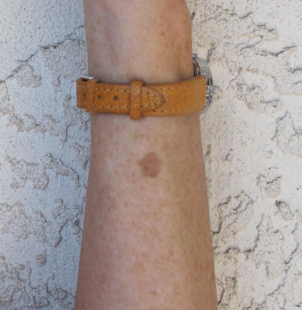
Sun-related pigmentation

Solar lentigo

A flat, sharply defined tan-to-brown patch—often called an age spot—on chronically sun-exposed skin such as the face, hands, forearms and shoulders.

Causes, types and diagnosis +

It reflects cumulative ultraviolet exposure and becomes more common with age. Facial lesions may develop a fingerprint-like or fine network pattern on dermoscopy. Irregular colour, shape or continuing enlargement can overlap with lentigo maligna, pigmented actinic keratosis or seborrhoeic keratosis and warrants assessment.

Treatment and outlook +

It is harmless and treatment is optional. Daily sun protection helps prevent further lesions. Cryotherapy, pigment lasers, intense pulsed light or selected topical agents may lighten confirmed benign lesions, but pigmentation can recur and treatment should not precede diagnostic certainty.

04

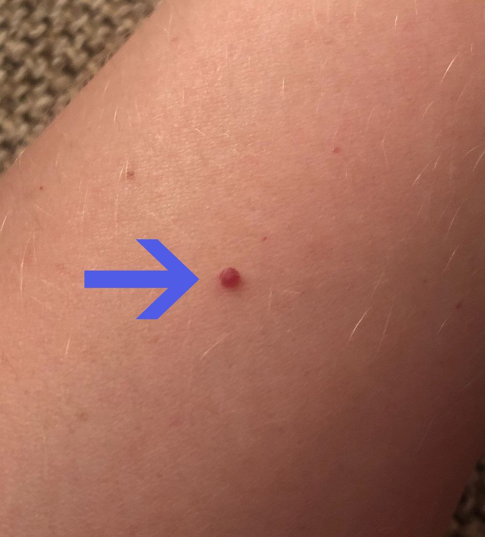
Benign blood-vessel growth

Cherry angioma

A small, round, bright-red to purple papule, usually on the trunk or limbs. Numbers commonly increase through adult life.

Appearance and diagnosis +

The cause is not fully understood; age and genetic tendency contribute. Dermoscopy typically shows well-defined red, purple or blue-red “lagoons.” A thrombosed angioma may turn dark and resemble melanoma, while a rapidly growing bleeding vascular lesion may be a pyogenic granuloma or, rarely, malignancy.

Treatment and outlook +

No treatment is needed. Electrocautery, vascular laser, cryotherapy or shave/curettage can remove lesions that bleed or are cosmetically troublesome. They remain benign, but new unexplained widespread vascular lesions should be discussed with a clinician.

05

Firm fibrous nodule

Dermatofibroma

A firm, usually small papule or nodule, most often on the lower legs. It may be pink, tan, brown or darker and often dimples when squeezed from the sides.

Causes, types and diagnosis +

It represents a benign fibrous reaction, sometimes following minor trauma or an insect bite. Dermoscopy often shows a central white scar-like area with a delicate peripheral pigment network. Larger, rapidly growing, ulcerated or atypical lesions need assessment because DFSP, melanoma and other tumours may mimic it.

Treatment and outlook +

Most need only reassurance. Complete excision provides diagnosis and treatment if painful, changing or uncertain, but leaves a scar and simple shave removal often recurs because the lesion extends into the dermis. Ordinary dermatofibromas do not metastasise.

06

Enlarged oil gland

Sebaceous hyperplasia

Small soft yellowish or skin-coloured papules, usually on the forehead, cheeks or nose, often with a central depression.

Causes and diagnosis +

Normal sebaceous glands enlarge, particularly with age, oily skin or immune-suppressing medicines. Dermoscopy may show yellow lobules arranged like a crown around a central opening, with vessels that do not cross the centre. A solitary firm, ulcerated or irregular lesion may need biopsy because BCC and sebaceous tumours can look similar.

Treatment and outlook +

It is harmless and does not require treatment. Electrosurgery, laser, cryotherapy, curettage or selected medical treatments may improve appearance, although recurrence or new lesions are common.

07

Keratin-filled lump

Epidermoid cyst

A slowly growing, mobile lump under the skin, often with a central pore. It contains keratin rather than “sebum” and can become inflamed or rupture.

Causes and diagnosis +

It forms when epidermal cells become trapped within the dermis, sometimes after trauma or from a blocked follicular opening. Diagnosis is usually clinical. Redness and tenderness may reflect sterile rupture rather than infection. An unusually firm, fixed, rapidly enlarging or recurrent mass may need imaging, biopsy or excision.

Treatment and outlook +

An asymptomatic cyst may be observed. Inflamed cysts may need anti-inflammatory treatment or drainage if fluctuant; antibiotics are reserved for genuine infection. Definitive treatment removes the intact cyst wall after inflammation settles. Recurrence is possible if the wall remains; malignant change is exceptionally rare.

08

Soft pedunculated growth

Skin tag

A small, soft, skin-coloured or brown growth on a stalk, commonly found on the neck, armpits, groin, eyelids and beneath the breasts.

Causes and diagnosis +

Skin tags are more common where skin rubs and in people with increasing age, obesity, pregnancy or insulin resistance. Diagnosis is usually straightforward. A pigmented, firm, ulcerated or atypical “tag” should be examined to exclude a naevus, seborrhoeic keratosis, neurofibroma or skin cancer.

Treatment and outlook +

They need no treatment. Snip excision, electrosurgery or cryotherapy can treat irritating lesions. Home cutting or tying is discouraged because of bleeding, infection and mistaken diagnosis. Treated tags do not usually return, but new ones may develop.

09

Common viral lesion

Viral wart

A rough papule caused by human papillomavirus (HPV). Common, flat, plantar, periungual and genital warts have different appearances and sites.

Spread and diagnosis +

Warts spread through direct contact or contaminated surfaces, particularly through damaged or wet skin. Dermoscopy may reveal interrupted skin lines and dotted or coiled vessels. On sun-damaged or immune-suppressed skin, a persistent “wart” may require biopsy to exclude SCC; genital lesions require a specific clinical assessment.

Treatment and outlook +

Many resolve through immune clearance, especially in children. Salicylic acid, cryotherapy and other clinician-directed options can help, but repeated treatment is often needed. Avoid picking and do not share razors or nail tools. Recalcitrant, painful, bleeding or rapidly changing lesions should be reviewed.

10

Benign fatty lump

Lipoma

A soft, rubbery, mobile lump beneath normal skin, commonly found on the trunk, shoulders, neck or limbs.

Causes and diagnosis +

The cause is often unknown and multiple lipomas can run in families. Most are diagnosed by examination; ultrasound or other imaging may be used when the lump is deep or atypical. A rapidly growing, painful, hard, fixed, deep or larger mass requires assessment to exclude another soft-tissue tumour.

Treatment and outlook +

Observation is suitable for a typical painless lipoma. Surgical excision may be chosen for symptoms, growth, diagnostic uncertainty or appearance. Recurrence after complete removal is uncommon, and an ordinary lipoma does not transform into liposarcoma.

How to use these images

Appearance is only one clue

These are representative clinical photographs, not diagnostic templates. Dermoscopy is especially useful for pigmented, vascular and keratotic lesions; for deeper lumps such as cysts and lipomas, palpation, ultrasound or pathology may be more informative. Any atypical or changing lesion should be assessed in person.

Less common, still important

Other rare skin cancers

Rare cancers can resemble a cyst, bruise, scar, red patch or harmless lump. They cannot be diagnosed reliably from appearance alone and commonly require biopsy, specialist pathology and multidisciplinary treatment planning.

01

Merkel cell carcinoma

A rare, fast-growing neuroendocrine cancer, often presenting as a painless red, pink, purple or skin-coloured firm nodule on sun-exposed skin. It is more common in older or immunosuppressed people and can spread early. Prompt biopsy, nodal assessment and specialist treatment—usually surgery with consideration of radiotherapy and, for advanced disease, immunotherapy—are important.

02

Sebaceous carcinoma

A cancer of oil-producing glands, most often around the eyelid, where it can imitate a persistent chalazion or eyelid inflammation. It may also occur elsewhere as a yellowish or pink nodule. Complete excision with careful margin assessment is usual; selected patients may need evaluation for Muir–Torre/Lynch syndrome.

03

Dermatofibrosarcoma protuberans

A slow-growing cancer arising in the deeper skin, often on the trunk or proximal limbs. It may begin as a firm scar-like plaque and later develop nodules. Spread to distant sites is uncommon, but local extensions can be wider than they appear. Wide excision or Mohs surgery is generally used to obtain clear margins.

04

Cutaneous angiosarcoma

An aggressive blood-vessel cancer that may resemble an enlarging bruise, purple-red patch or swelling, classically on the scalp or face of an older person. It can also occur after radiotherapy or chronic lymphoedema. Diagnosis needs a biopsy; treatment may combine surgery, radiotherapy and systemic therapy through a sarcoma team.

05

Kaposi sarcoma

A cancer caused by human herpesvirus 8 (HHV-8), often appearing as painless red, purple (violaceous), brown or nearly black spots, plaques or nodules. Lesions may occur on the skin or inside the mouth and can be mistaken for bruises. HIV-associated Kaposi sarcoma is an AIDS-defining illness, but Kaposi sarcoma also occurs in people taking immune-suppressing medicines and in classic or endemic forms. Diagnosis normally requires biopsy and assessment for disease elsewhere. For HIV-associated disease, antiretroviral therapy is central; local or systemic cancer treatment may also be required.

06

Atypical fibroxanthoma and pleomorphic dermal sarcoma

Atypical fibroxanthoma (AFX) usually presents as a rapidly growing pink-red, sometimes ulcerated nodule on heavily sun-damaged skin of an older person, especially the scalp, face, ears or neck. It is a superficial tumour with a low metastatic risk after complete removal, but diagnosis requires pathology and immunohistochemistry to exclude look-alikes. Pleomorphic dermal sarcoma (PDS) is its deeper, more aggressive counterpart and has greater risks of recurrence and spread. Complete margin-controlled surgery and follow-up are important.

07

Adnexal and other cancers

Rare tumours can arise from sweat glands, hair follicles or other skin structures; examples include microcystic adnexal carcinoma and porocarcinoma. Primary cutaneous lymphomas and skin metastases from internal cancers also occur. Their appearances overlap with common lesions, so accurate classification by pathology is essential before treatment.

Arrange reviewA new rapidly enlarging firm nodule, an unexplained purple or bruise-like patch that persists or spreads, a persistent “chalazion,” or a lesion associated with pain, numbness or nearby swollen glands should be medically assessed. Multiple violaceous skin or mouth lesions—particularly in a person with HIV or immune suppression—need prompt review.

Professional examination and digital monitoring

Skin checks with FotoFinder

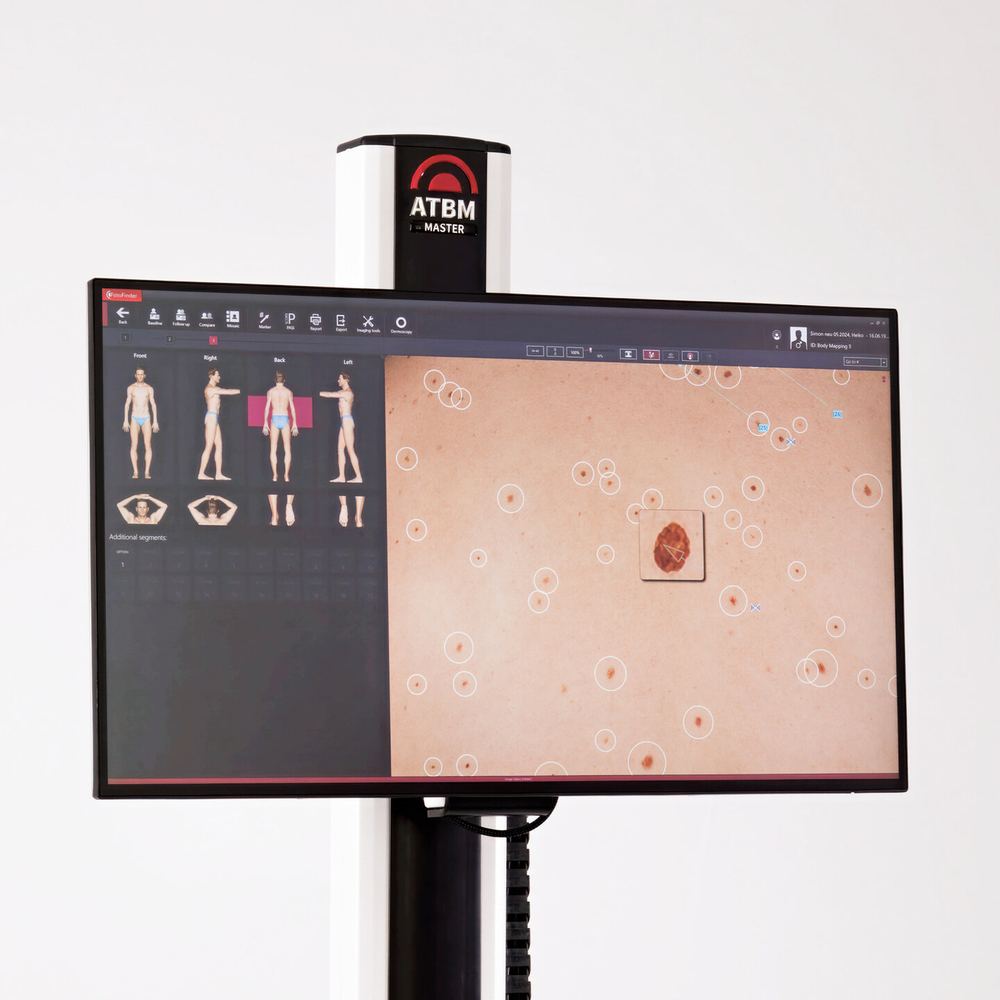
A professional skin check examines the whole skin surface—not only the spot that first caused concern. FotoFinder can add standardised total-body photography and dermoscopic imaging, creating a visual baseline that can be compared carefully at future visits.

Photography and artificial intelligence support the clinician. They do not diagnose cancer, replace examination or remove the need for biopsy when a lesion is suspicious.

FotoFinder ATBM master system displaying total-body mapping and computer-assisted identification of skin lesions
The FotoFinder ATBM master systemThe imaging station combines standardised total-body photography, lesion mapping and close-up digital dermoscopy. The display shown demonstrates how lesions can be catalogued for structured clinical review and later comparison.
Image © FotoFinder Systems GmbH
Allow45minutes

What to expect at your appointment

A thorough, clinician-led skin check

Please allow approximately 45 minutes for photography, a complete clinical examination, discussion of the findings and treatment planning where required. The appointment is designed to assess your whole skin surface—not only one lesion.

01

Nurse preparation and imaging

The practice nurse will call you in, explain the process and take standardised full-body photographs with FotoFinder. Dermoscopic images of selected lesions are also recorded.

02

Personal risk assessment

Dr Mikhail will ask about previous skin checks, any personal or family history of skin cancer, sun exposure and sunburn, immune suppression, changing lesions and the preventive measures you currently use.

03

Image review and examination

The recorded images are reviewed, followed by a systematic full-skin examination. A handheld dermatoscope is used to reassess documented lesions and examine any additional areas, helping ensure that clinically important lesions have not been missed.

04

Findings and a clear plan

You will discuss the findings, what they mean and the most appropriate next step. There is time to ask questions and, if treatment is needed, to understand the proposed procedure and follow-up.

After the examination

What may happen next?

No concerning lesions

Reassurance and a personalised prevention plan are provided. Depending on your risk, a routine skin check may be recommended in approximately 1–2 years.

A lesion needs monitoring

If a lesion is not clearly suspicious but remains uncertain, repeat clinical and dermoscopic imaging may be offered in about 2–3 months to assess for meaningful change.

A suspicious lesion is identified

When there is significant concern for a neoplastic process, a biopsy or complete removal may be recommended. The options, timing, pathology and treatment plan will be discussed with you.

01

Establish a baseline

Standardised photographs systematically record the skin surface. Selected moles and lesions can also be saved as magnified dermoscopic images.

02

Compare over time

At follow-up, current images are aligned with earlier images. This makes side-by-side comparison more consistent than relying on memory or isolated photographs.

03

Find what is new

Body-mapping software can highlight a lesion that was not visible at the previous visit—important because many melanomas arise as new lesions rather than within a longstanding mole.

04

Detect subtle change

Changes in size, shape, colour or dermoscopic structure can be identified and assessed. A lesion that looks only mildly unusual at one visit may become more significant when genuine evolution is demonstrated.

Why longitudinal imaging helps

A patient-specific “map” of your skin

  • Better recognition of change: the clinician can compare the same body area and lesion across different dates.
  • Detection of new lesions: useful when there are many moles and it is difficult to remember which have always been present.
  • More precise dermoscopic follow-up: selected equivocal lesions can be monitored for structural change when immediate biopsy is not indicated.
  • A more reproducible record: standardised positioning and image capture reduce variation between visits.
  • Supports targeted decisions: documented stability may be reassuring, while meaningful evolution can prompt biopsy or excision.
AI-assisted

What does the inbuilt AI do?

FotoFinder’s computer-assisted analysis evaluates visual and dermoscopic features and can flag or score lesions that warrant closer attention. It acts as an additional decision-support tool for the trained clinician, alongside history, examination, dermoscopy and comparison with previous images.

Important limitationA low-risk AI assessment cannot rule out melanoma, and a high-risk result does not prove cancer. Some melanomas are subtle or non-pigmented; suspicious lesions still require clinical judgement and, where indicated, histopathology.

How often should I have a skin check?

The interval should match your individual risk

Australia does not have one fixed screening interval for every adult. A clinician should assess your history, skin type, sun exposure, number and type of moles, previous cancers, family history and immune status.

Higher riskClinical skin examination is commonly recommended every 6–12 months, with photography where helpful.
After skin cancerFollow-up is personalised. Checks may be every 3–6 months after a high-risk cancer and around yearly after a low-risk cancer, depending on the treating clinician’s plan.
Lower or average riskThere is no universal timetable. Discuss your risk with your GP or skin cancer clinician and continue regular self-examination.
A concerning lesionDo not wait for a routine appointment. Arrange prompt review for a new, changing, bleeding, painful or non-healing lesion.

Who needs closer surveillance?

Factors that increase melanoma or skin-cancer risk

  • Previous melanoma, BCC, SCC or numerous actinic keratoses
  • A first-degree relative with melanoma, especially multiple affected relatives
  • Many moles, atypical/dysplastic moles, or a very large congenital naevus
  • Fair skin, light eyes, red or fair hair, freckles, or skin that burns readily
  • Repeated sunburn—particularly blistering sunburn in childhood—or high cumulative UV exposure
  • Outdoor work, outdoor recreation, solarium use, or residence in a high-UV environment
  • Immune suppression, including organ transplantation or immune-suppressing medicines
  • Older age and male sex, although melanoma can occur at any age and in all skin tones
  • Genetic cancer syndromes or a clinician-assessed strong personal/family risk profile

Sources: FotoFinder total-body mapping, Cancer Council Australia early detection statement and follow-up guidance.

Protect today · reduce future risk

Skin cancer prevention

Most skin cancers are linked to ultraviolet (UV) exposure accumulated over many years. Good protection is not one product—it is a routine that combines clothing, sunscreen, a protective hat, shade and sunglasses whenever the UV Index is forecast to reach 3 or above.

UV cannot be seen or felt, and cool, cloudy or windy weather can still have damaging UV levels. Check the day’s UV forecast, not the temperature.

01

Slip

Cover skin

Choose long sleeves, collars and longer garments. Densely woven, loose-fitting fabric or clothing labelled UPF 50+ gives the best protection.
02

Slop

Use sunscreen

Apply SPF 50 or SPF 50+, broad-spectrum and water-resistant sunscreen generously to every area clothing does not cover.
03

Slap

Wear a shady hat

A broad-brimmed, bucket or legionnaire hat should shade the face, ears and neck. A cap or visor leaves important areas exposed.
04

Seek

Use shade

Plan outdoor activity and breaks around shade, especially near midday. Reflected and scattered UV still reaches skin under shade.
05

Slide

Protect the eyes

Choose close-fitting wraparound sunglasses labelled AS/NZS 1067.1, lens category 2, 3 or 4—not fashion spectacles.

Choosing sunscreen

Four things to find on the label

SPF 50 or 50+High UVB protection when applied at the tested amount.
Broad-spectrumProtection from both UVA and UVB radiation.
Water-resistantImportant for swimming, sport, sweating and long outdoor days.
Australian compliantUse an in-date sunscreen supplied for Australia; follow its label and store below 30°C.

The best formulation is one you will use properly. Lotion, cream, gel, spray, mineral or organic-filter products can all be suitable if they meet the criteria above. For sensitive skin, try a fragrance-free product or a small test area. Sprays must still be applied generously and rubbed in—avoid inhalation and do not rely on a light mist.

The teaspoon rule

About 35 mL for an adult full body

Use approximately one teaspoon for each area:

  • face, neck and ears
  • each arm
  • each leg
  • front of the body
  • back of the body

Often missed: ears, lips, bald scalp and hairline, sides of the neck, backs of hands, feet and around swimwear edges.

Use it correctly

Match your routine to the day

Sunscreen wears, rubs and washes off. A morning application is useful, but it is not enough for a full outdoor day.

Everyday routine

Apply before leaving home

  • Check when the UV will be 3 or above.
  • Apply to clean, dry exposed skin 20 minutes before going outdoors.
  • Include it before make-up; let sunscreen settle first.
  • Reapply if you remain outdoors, sweat or wipe it off.

Prolonged exposure

Build in reapplication

  • Use all five forms of protection.
  • Reapply sunscreen every two hours outdoors.
  • Reapply sooner after heavy sweating or rubbing.
  • Plan shade and breaks; sunscreen should not extend time in the sun.

Beach, pool or boating

Water resistance is not waterproof

  • Apply water-resistant SPF 50+ before dressing or swimwear.
  • Reapply every two hours and immediately after swimming, sweating or towel-drying.
  • Wear a rash vest or UPF 50+ swimwear and a secure hat.
  • Use shade; water and sand can reflect UV.

Medication-assisted prevention

Nicotinamide (niacinamide): useful for selected high-risk patients

Oral nicotinamide is the amide form of vitamin B3. In a 12-month Australian trial involving people who had at least two non-melanoma skin cancers in the previous five years, 500 mg twice daily reduced the rate of new basal and squamous cell cancers by about 23% while treatment continued. The benefit was not maintained after it was stopped.

Who may benefit?People with repeated BCCs or SCCs, extensive actinic damage or another clearly high-risk history—after discussion with their treating clinician.
Important limitsIt has not been proven to prevent melanoma, does not prevent sunburn and never replaces sun protection or skin checks. Evidence in organ-transplant recipients has not shown the same benefit.
Use the correct formChoose nicotinamide/niacinamide—not nicotinic acid (niacin), which has different effects and can cause flushing. Cosmetic niacinamide cream is not a substitute for the studied oral regimen.
Check firstDiscuss pregnancy or breastfeeding, liver or kidney disease, diabetes, low platelets and medicines with a doctor or pharmacist. Side effects can include nausea, headache or diarrhoea.
No tablet or sunscreen provides complete protection.Avoid tanning and solariums, protect children early, do not deliberately seek extra UV for vitamin D, and continue self-checks and risk-based professional skin examinations.

Sources: Cancer Council Australia SunSmart guidance, sunscreen application guidance, eye protection guidance and the ONTRAC nicotinamide trial.

Build a simple routine

Check your skin and know your normal

Use a well-lit room, a full-length mirror and a hand mirror. Ask someone you trust to help with your scalp and back.

  1. 01

    Look everywhere

    Face, ears, scalp, trunk, arms, hands, legs, soles, between toes, nails and genital skin.

  2. 02

    Compare

    Notice a spot that looks unlike the others—the “ugly duckling”—or a lesion that has changed.

  3. 03

    Record

    Note the date and location. A clear photograph beside a ruler can help document change.

  4. 04

    Act

    If you are concerned, book a medical examination. Do not rely on an app or image comparison.

A useful memory aid for pigmented lesions

ABCDE of melanoma

Not every melanoma follows this rule. Nodular and amelanotic melanoma may be symmetrical or have little pigment.

A
Asymmetry
One half differs from the other
B
Border
Irregular or poorly defined edge
C
Colour
More than one colour or uneven colour
D
Diameter
Often over 6 mm, but may be smaller
E
Evolving
Changing size, shape, colour or symptoms

For fast-growing or nodular melanoma

Remember EFG

EElevated above the skin
FFirm to touch
GGrowing progressively—often over weeks

Photograph consistently

Use the same room, lighting, camera distance and body position. Include a ruler without covering the lesion, keep the original dated file and avoid filters or digital zoom. Photographs document change but do not replace dermoscopy.

Monthly routine: face and scalp → front and sides → arms and hands → back and buttocks → legs, feet, soles and between toes → nails and genital skin. Ask a partner to check areas you cannot see.

Easy-to-miss locations

Skin cancer is not always in an obvious place

Include these sites in self-checks and professional examinations—particularly when a symptom persists without a clear explanation.

01

Scalp & behind ears

Use a comb or hairdryer in sections; ask a partner for help. Bald and thinning scalps receive substantial UV.

02

Palms, soles & between toes

Acral melanoma can occur in any skin tone and is not necessarily related to sun exposure.

03

Nails

Look for a new or widening pigment band, pigment spreading onto surrounding skin, nail splitting or a persistent lesion beneath the nail.

04

Eyelids & lips

Persistent lash loss, ulceration, distortion of the lid margin, or a scaly/firm lip lesion warrants examination.

05

Genital & perianal skin

New pigment, a persistent sore, lump, bleeding or unexplained itch should be assessed sensitively and promptly.

06

Mouth & mucosa

Persistent pigmented patches, ulcers, bleeding or lumps require dental or medical review; mucosal melanoma is rare but serious.

What happens next?

From suspicious lesion to a clear plan

Your clinician chooses the safest diagnostic route based on the lesion and your health. Sometimes the whole lesion is removed at the first procedure; sometimes a sample is taken first.

  1. 1

    Assessment

    History, whole-skin context, visual examination, palpation and dermoscopy.

  2. 2

    Biopsy or excision

    Shave, punch, incisional or complete excision under local anaesthesia, selected to answer the clinical question.

  3. 3

    Histopathology

    A pathologist identifies the diagnosis and reports features such as subtype, depth and margins. Timing varies; ask when and how results will be delivered.

  4. 4

    Treatment plan

    Options may include observation, wider excision, margin-controlled/Mohs surgery, topical or destructive treatment, radiotherapy or specialist referral.

  5. 5

    Follow-up

    Wound review, pathology discussion, scar care and risk-based surveillance for recurrence or new cancers.

Common treatments

Excision
Removes the lesion with a planned margin; stitches, a local flap or skin graft may close the wound.
Curettage/cautery
Scrapes and seals selected superficial lesions; heals like a graze.
Cryotherapy
Freezes suitable lesions; blistering, crusting and pigment change can occur.
Topical therapy/PDT
Treats selected superficial cancers or fields; redness and crusting are expected and require a prescribed plan.
Mohs surgery
Examines margins in stages while preserving healthy tissue; useful for selected high-risk sites and tumours.

Recovery and wound safety

  • Follow the specific dressing and activity instructions from your treating team.
  • Keep early wounds protected; return to exercise, work and swimming depends on site, closure and treatment.
  • Scars commonly remain pink and firm before softening over 6–18 months.
  • Seek advice for increasing pain, spreading redness, pus, fever, wound separation or bleeding that does not stop with firm pressure.
  • Do not assume “clear margins” means no future checks are needed.

Personalised surveillance

Groups needing particular care

Risk is cumulative. People with several factors often benefit most from a documented surveillance plan, total-body photography and dermoscopy.

Previous melanoma or repeated cancers

Risk of another primary cancer is increased; follow the schedule set by the treating team.

Transplant or immune suppression

SCC risk may rise markedly and cancers can behave more aggressively. Medication changes must be specialist-led.

HIV or haematological disease

Immune status changes the spectrum and behaviour of skin cancer, including Kaposi sarcoma.

Strong family or genetic risk

Multiple relatives, young-onset melanoma or known predisposition syndromes may justify specialist surveillance or genetic counselling.

Numerous or atypical moles

The ugly-duckling approach plus total-body and sequential dermoscopic photography can help identify meaningful change.

High UV or treatment exposure

Outdoor work, solariums, previous radiotherapy or PUVA add risk; workplace protection and long-term surveillance matter.

If a lesion worries you

Arrange an in-person skin examination

Contact your GP, skin cancer doctor or dermatologist. Tell the receptionist if the lesion is rapidly growing, bleeding repeatedly or has changed noticeably.

Urgent medical help

Severe bleeding or acute illness?

Apply firm continuous pressure to bleeding. In Australia, call 000 for uncontrolled bleeding or a medical emergency. For health advice, call healthdirect on 1800 022 222.

Find an Australian health service

About this guide

Education, not diagnosis

This guide is written for the public and uses selected examples. Skin cancers have many appearances across different skin tones and body sites. A normal-looking photograph does not rule out cancer, and a worrying-looking photograph does not prove it.

Clinical information is based on Australian patient guidance and recognised dermatology references. Treatment choices depend on pathology, site, size, depth, patient health and clinician assessment.

Please note that this guide is general education only. Photographs cannot confirm a diagnosis, and information here should not replace an in-person skin examination or professional medical advice.

Noticed a spot that concerns you?

Book a skin check with our clinic and get a clear, professional assessment.

Book your skin check
Shopping Cart
×